

Murray Wood: Artemis is what the world needed this week
Murray Wood says the Artemis II moon mission is sorely needed right now. Wood says you'd never know from looking at the ...
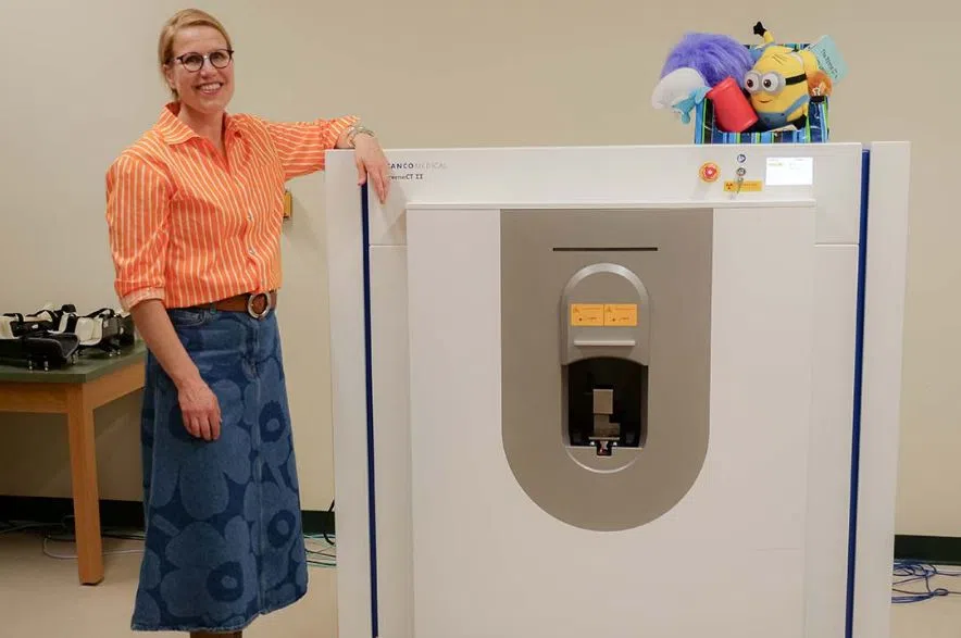
A team of University of Saskatchewan researchers will be able to study more about children’s bone health without breaking any, with the help of advanced imaging tools.
Traditionally, the only way to gauge the strength of our bones would be to break them.
Read more:
The new technological tools will allow researchers “to capture insights into bone strength, all while keeping them intact,” said the press release from USask.
“It’s challenging to actually study bone strength, but understanding bone strength and bone growth is necessary to develop therapies that can protect people from fractures throughout their life,” said Dr. Saija Kontulainen (PhD), professor and associate dean of research in the College of Kinesiology.
Kontulainen and her colleagues, Dr. J.D. Johnston (PhD) from USask’s College of Engineering and Dr. Munier Nour (MD) from USask’s College of Medicine, are conducting a first-of-its-kind pediatric bone study to understand what makes bones fragile.
“The findings of this project will help inform new therapies that can optimize bone strength development in children and youth who are at risk of bone fragility and fracture throughout their lives,” noted the university.
The Canada Foundation for Innovation’s John R. Evans Leaders Fund contributed to the university securing this improved imaging tool.
“The scanner is a second-generation, high-resolution peripheral quantitative computed tomography (HR-pQCT-II) and is a non-invasive way for us to look at the micro-architecture of the bone and the properties of the surrounding muscle,” she said.
The statement described the scanner as “powerful” and “quite compact.”
Due to the advanced imaging technology, the team of researchers can run engineering simulations to pinpoint how much force it takes to break a bone, using detailed images of the inside of the bone and the surrounding structures, said the release.
These details can help the research team to develop “new evidence-based approaches for bone health assessments and develop therapies to monitor fracture-prone populations, such as children with Type 1 diabetes and those diagnosed with autism spectrum disorder (ASD).”
“We also gain new insights into what influences bone development. This is all exciting because it offers monitoring of bone tissue in growing and aging skeleton, and the impact of therapies across the lifespan,” she said.
Kontulainen said that bone health among children with Type 1 diabetes and children with ASD is often overlooked and poorly understood, but healthy bones are crucial for longevity.
A better understanding of bone development can help inform therapies, including non-pharmaceutical interventions like lifestyle modifications and physical activity, added the university.
According to Kontulainen, early intervention can optimize bone strength and increase quality of life into older adulthood.
Kontulainen said that the new scanner is attracting interest from researchers and trainees across campus who need high-resolution images for their clinical and foundational studies.
“This scanner can be used for so many research projects and clinical studies across all ages, across campus,” she said.
“Having these kinds of advanced research tools available for use at the university benefits many populations, researchers and trainees to collaborate, including national and international studies. That is why continued investment into research infrastructure like this is so important and impactful.”
Read more:


")